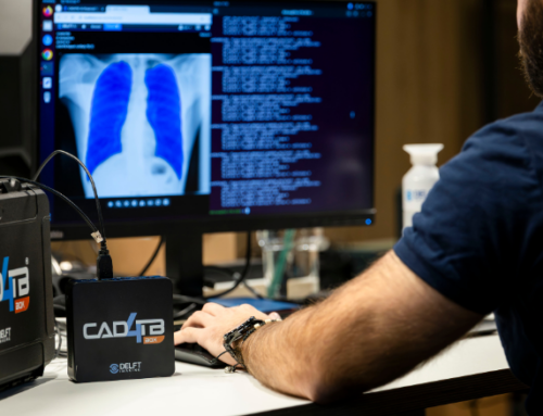
The 2024 Delft Imaging webinar hosted Prof. Rodney Ehrlich, a Senior Research Scholar from the Division of Occupational Medicine at the University of Cape Town. His presentation, titled “Overcoming Obstacles in Implementing Computer-Aided Detection for Silicosis & TB,” provided an in-depth look into the challenges and opportunities presented by AI-enhanced medical imaging solutions, particularly in the context of silicosis and tuberculosis (TB) detection.
The Challenges of Diagnosing Silicosis
Silicosis poses a unique diagnostic challenge compared to other lung diseases. As Prof. Ehrlich noted, only a third of silicosis cases are visible on chest X-rays (CXR) compared to autopsy findings. This discrepancy underscores the need for advanced X-ray software for silicosis that can distinguish between different radiological phenotypes and differentiate silicosis from other lung conditions like TB.
“Silicosis is much more sensitive. When I say sensitive, I mean sensitive to being missed or misdiagnosed with poor quality chest x-rays. Now, this was particularly a problem with analog films and it should be disappearing with digital radiology, but it seems not necessarily to be the case,” he stated.
“The reason is when you have a symmetrical nodular pattern of nodules in the X-ray, it can be very difficult to distinguish from normal broncho vascular markings. This is not the case, for example, if you have a mass in one lung or you have a pleural effusion. Sharp contrast is needed,” he continued
“And also, you need to read these x-rays on a high standard monitor screen. Otherwise, you’re going to make mistakes. And, you know, obviously in remote settings, this becomes a big issue.”
AI in Medical Imaging: Bridging the Gap
The integration of AI in medical imaging is transforming how we approach the diagnosis of complex diseases. Prof. Ehrlich’s discussion shed light on the promise of silicosis detection algorithms that can enhance the accuracy of screening processes. These algorithms are crucial for distinguishing between silicosis, TB, and combined conditions like silicotuberculosis.
Overcoming Technical and Operational Hurdles
Despite the potential of AI-enhanced analysis, several obstacles remain. Prof. Ehrlich pointed out the variability in performance among different software vendors and the need for transparency in the source of training images, protocols, and validation outcomes. This variability necessitates continual software improvements and clinical research to develop reliable criteria for clinical validation and the choice of appropriate thresholds based on specific use cases.
The Role of X-Ray Software for TB Detection
In regions with high TB burdens, the overlap between TB and silicosis is significant. Prof. Ehrlich stressed the importance of utilizing X-ray software for TB detection in conjunction with silicosis screening.
“The problem with silicosis is no gold standard. Unlike TB, there’s no high accuracy, while gold standard would have been a high accuracy reference standard that we’ve heard about, other than autopsy,” Prof. Ehrlich pointed out. “So CT could play an important role here. And there’s no question that CT is more sensitive in picking up silicosis, but it also identifies specific features.
This dual approach is essential in mining areas where both diseases are prevalent. Accurately identifying and differentiating between TB and silicosis on CXRs can significantly impact public health strategies and patient management.
Implementing Effective Screening Programs
For CAD systems to be effective, there must be a shift from merely adopting new technology to integrating it into programmatic frameworks. This involves educating users about the practical applications of silicosis imaging solutions and ensuring that these tools are user-friendly and align with clinical workflows. Prof. Ehrlich called for comprehensive operations research to evaluate the impact of CAD on compensation systems, fitness for work policies, and overall cost-efficiency.
Conclusion
Prof. Ehrlich’s webinar highlighted AI’s potential in easing the detection and management of silicosis and TB.
“We need more clinical research, and particularly, I think, for silicosis, something to match the article by Kahn, 2017, computer-aided reading of tuberculosis chest radiography, moving the search agenda towards a foreign policy in which they set out a type of study design and everything to do with study and ethics, in other words, a sort of practical guide or set of criteria by which clinical validation studies should be judged. The problem of threshold shifts needs an algorithm for the local users; it’s a question the user needs to ask: what threshold should I use?” Prof. Ehrlich asked.
According to Prof. Ehrlich, by addressing the technical and operational challenges, AI-driven X-ray software for silicosis and TB can significantly improve screening accuracy and efficiency. “Operations research, we heard from my colleague, that is needed. That’s not well-funded. It seems that some funders’ eyes glaze over when you use the word operations research. We’re talking about everything that we’ve heard about today, side-by-side comparisons with actual doctors and radiographers on the ground, ease of use, connectivity, agreement with readers, but I don’t mean validation, I mean agreement with the doctors using it, because if the doctors and the clinical assistants, clinical radiographers see things they don’t agree with, they’re going to lose faith in the CAD. And, of course, the outcomes and efficiency of its compensation outcomes, management, fitness for work, or screening for TB in relation to cost.”
As research and technology continue to advance, the integration of AI-enhanced analysis will play a pivotal role in combating these pervasive occupational lung diseases.


